

.png?width=256&height=256&name=customer-service(1).png)

.png?width=64&height=64&name=calendrier(1).png)
.png?width=64&height=64&name=communique-de-presse(1).png)
.png?width=64&height=64&name=livre(1).png)
.png?width=64&height=64&name=blog(2).png)


Pathology Informatics 2025
Join Tribun Health at Pathology Informatics in Ann Arbor.
From 19/05/2025 to 22/05/2025
Ann Arbor
B1
Congresses
Pathology Informatics 2025
Join Tribun Health at Pathology Informatics in Ann Arbor.
From 19/05/2025 to 22/05/2025
Ann Arbor
B1
Congresses

Canada's Inaugural DP & Precision Diagnostics Summit 2025
Join Tribun Health at Canada's Inaugural Digital Pathology & Precision Diagnostics 2 Day Summit in Montreal.
From 08/05/2025 to 09/05/2025
Montreal
Congresses

USCAP 2025
Join Tribun Health at USCAP in Boston.
From 24/03/2025 to 26/03/2025
Boston
Booth n°936
Congresses
JIAP 2025
Join Tribun Health at JIAP (1st AI and Pathology Day) in Paris.
From 13/03/2025 to 13/03/2025
Paris
Booth n°4
Congresses
HIMSS 2025
Join Tribun Health at HIMSS 2025 in Las Vegas.
From 03/03/2025 to 06/03/2025
Las Vegas
Booth C3102-17 in the Startup Park Pavilion
Congresses
CAP 2024
Join Tribun Health at the CAP-ACP 2024 Annual Scientific Meeting 2024.
From 05/07/2024 to 06/07/2024
Halifax - Marriott Harbourfront Hotel
Booth #13
Congresses

ECP 2024
Join Tribun Health at the European Congress of Pathology 2024.
From 07/09/2024 to 11/09/2024
Florence - Fortezza da Basso
Booth #93
Congresses

Carrefour Pathologie 2024
Join Tribun Health at Carrefour Pathologie 2024.
From 20/11/2024 to 22/11/2024
Paris, France
Booth #27
Congresses

DP & AI Congress Europe 2024
Join Tribun Health at the DP & AI Congress Europe 2024.
From 11/12/2024 to 12/12/2024
London, UK
Booth #45
N°1
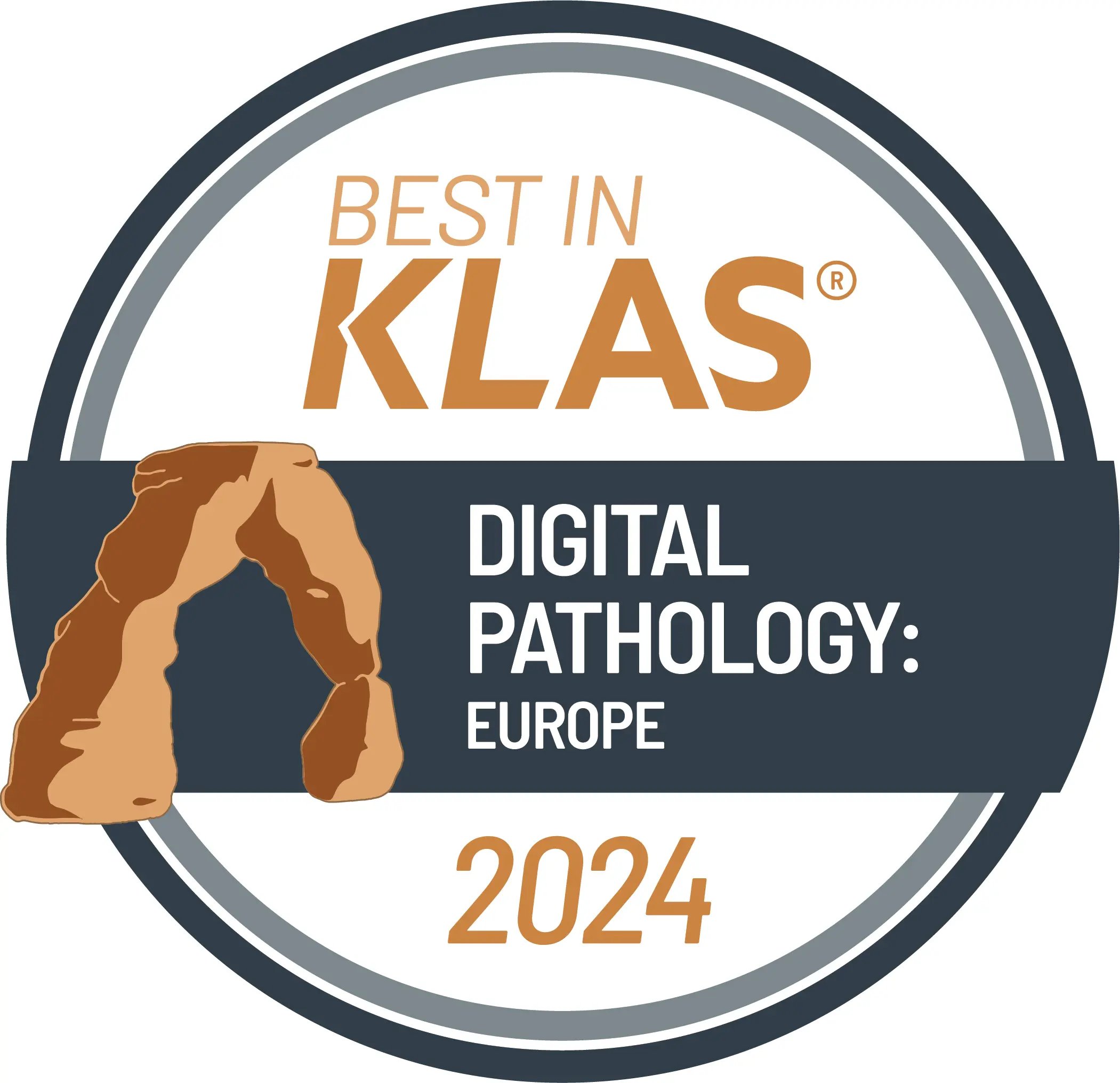
DigPath Platform I 3x Best in KLAS Winner
+200
Customers, including diagnostic & pharma labs
+50
Specialists forging reliable solutions

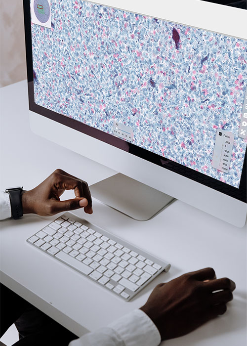
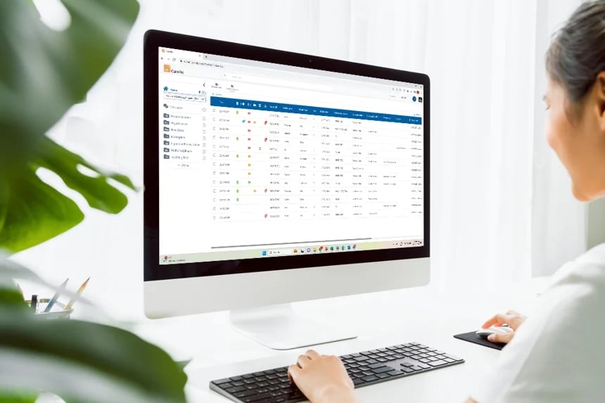
Discover Tribun Health's end-to-end platform, comprising all the modules we offer: MacroCam, CaloPix Archive, AI Apps, CaloPix® and TeleSlide. Together, they meet the end-to-end digitization needs of pathology departments, and integrate seamlessly into pathologists' workflows. At the heart of the digital pathology workflow is our Image Management System (IMS), CaloPix®.