


.png?width=256&height=256&name=customer-service(1).png)
.webp)

.png?width=64&height=64&name=calendrier(1).png)
.png?width=64&height=64&name=communique-de-presse(1).png)
.png?width=64&height=64&name=livre(1).png)
.png?width=64&height=64&name=blog(2).png)


Macroscopy: A Key Step in Diagnosis
Macroscopy—also known as grossing—is often overlooked, but it’s the first and most critical step in the diagnostic chain. With digital innovation transforming pathology, macroscopy is evolving from a manual, error-prone task to a streamlined, traceable, and collaborative process.
Traditionally performed with handwritten notes and limited traceability, this visual examination of surgical specimens has long been essential for identifying which areas need microscopic analysis. But in a world where labs must deliver results faster, more securely, and with fewer resources, modernizing macroscopy isn’t optional—it’s urgent.
Let’s explore why macroscopy matters, where traditional workflows fall short, and how digital tools are transforming this foundational step.
Why Macroscopy Matters in Pathology
In healthcare, precision is non-negotiable. And in pathology, precision begins long before a specimen reaches the slide image review.
Here’s how macroscopy shapes diagnosis and why its modernization is essential:
1. The First Line of Defense in Diagnosis
Macroscopy is where the diagnostic process begins. It’s a visual and tactile assessment that determines which parts of the specimen are examined under the microscope. If a tumor margin or suspicious lesion is overlooked here, it won’t be captured later—no matter how advanced the imaging or analysis tools.
Even the best microscopic interpretation can’t recover what was never sampled.
2. The Blueprint for Sampling and Staging
Macroscopic assessment guides tissue sampling and plays a critical role in cancer staging. For example, only macroscopy can reliably distinguish between continuous tumour invasion and separate tumour foci. These details shape clinical decisions—from prognosis to therapy selection.
3. A One-Time Opportunity
Specimens are only handled once. After they’re sectioned or discarded, their spatial and anatomical context is lost. If something is missed during macroscopy, there’s no way to go back. That makes this step not just important—but irreplaceable.
4. Direct Impact on Treatment Plans
Macroscopic findings can have immediate clinical consequences. Identifying subtle but high-risk features—like a non-seminomatous component in a testicular tumour—can change a patient’s entire care plan. These aren’t academic observations. They’re life-altering calls.
5. The First Chapter of the Pathology Report
Macroscopy isn’t just the first step chronologically—it sets the tone for everything that follows. It includes visible details like the size, color, consistency, and margins of the specimen. This information gives pathologists and clinicians essential context to interpret the histological findings that follow.
The Limits of Traditional Grossing
Conventional macroscopy methods present real challenges in today’s clinical environment:
-
Inconsistent traceability, especially when performed by someone other than the reporting pathologist
-
Manual data entry errors, from specimen ID to documentation
-
Fragmented communication between lab technicians, pathology assistants, pathologists, and surgeons
These problems are amplified in time-sensitive scenarios like frozen section exams, where rapid diagnostic input is needed to guide a surgical procedure in real time. Delays or miscommunications here can directly affect patient safety.
In an era when labs are under pressure to boost productivity, reduce errors, and protect data integrity, traditional macroscopy methods are no longer enough.
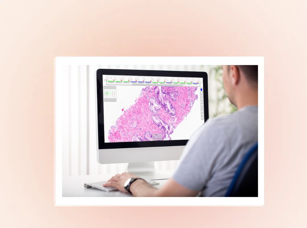
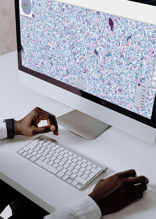
How Digital Tools Are Modernizing Macroscopy
The shift to digital macroscopy brings major advantages. By integrating with the digital pathology workflow, these tools allow for:
-
High-resolution image capture
-
Live annotations and measurements
-
Automated links to LIS and patient records
-
Secure remote collaboration and review
The result? Greater accuracy, better coordination across teams, and dramatically improved traceability—from the lab bench to the clinical report.

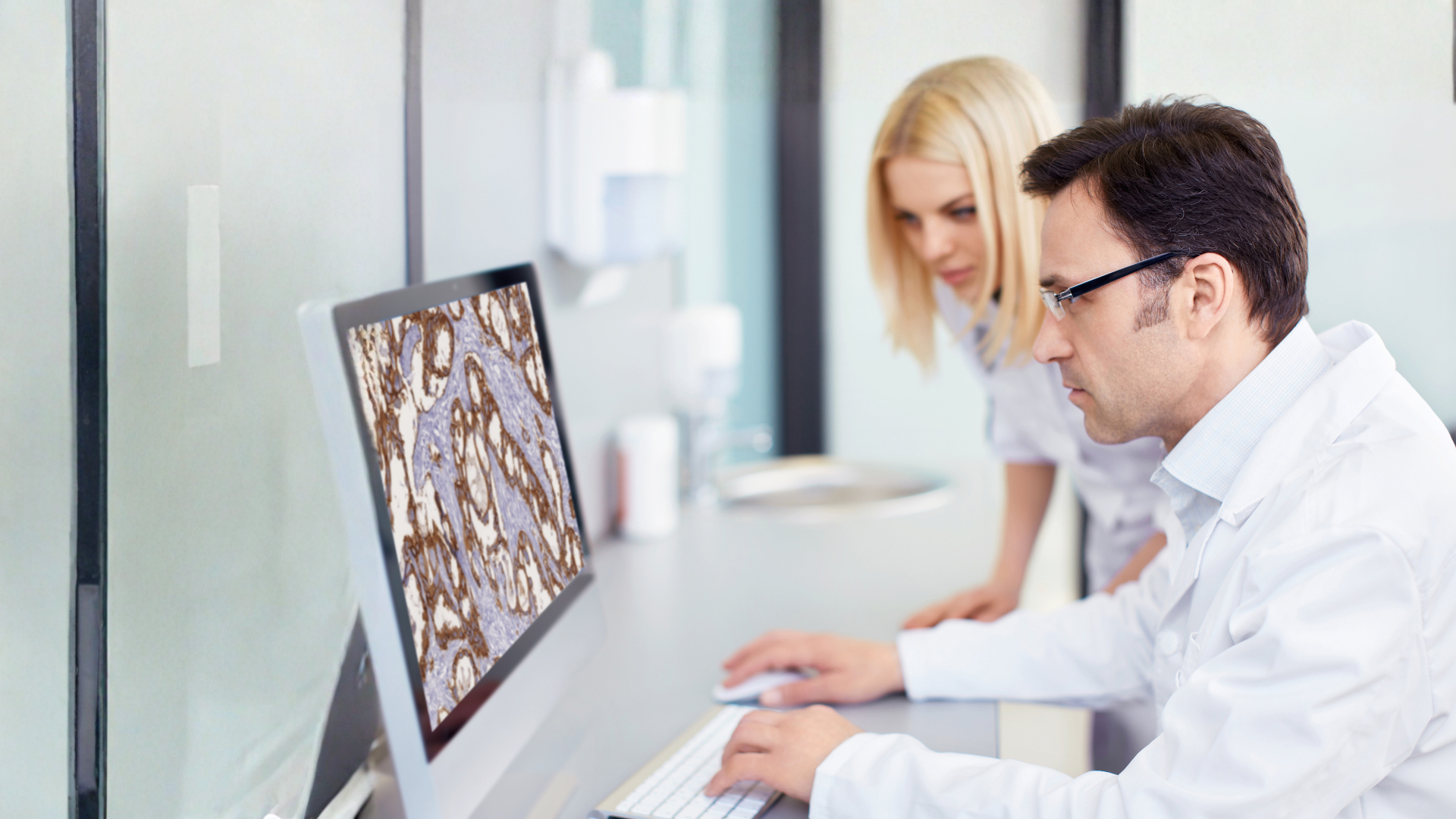
MacroCam: A Real-World Example
Digital macroscopy isn’t theoretical—it’s here. Tribun Health’s MacroCam is a high-definition imaging solution designed to simplify and standardize macroscopy.
With hands-free operation via footswitch, a built-in barcode scanner for traceability, and seamless integration with the LIS and CaloPix® image management platform, MacroCam supports fast, intuitive workflows.
Whether used for routine macroscopy or live frozen section collaboration, it empowers labs to:
-
Capture crisp, high-resolution images—even for small or fragile specimens
-
Annotate and measure images directly within the software
-
Share macroscopy results in real time across clinical sites
-
Maintain full traceability and audit readiness
-
Train junior pathologists and residents more effectively
In frozen section scenarios, MacroCam allows geographically dispersed teams to collaborate live—enabling real-time surgical decision support and greater confidence.
Benefits of Digital Macroscopy for Laboratories
Labs that adopt digital macroscopy solutions like MacroCam benefit from:
✅ Improved image quality, with HD resolution
✅ Faster workflows, reducing bottlenecks in specimen handling
✅ Complete traceability, from acquisition to report
✅ Enhanced collaboration, across technicians, pathologists, and clinicians
Conclusion: Getting It Right the First Time
Macroscopy may be the first step in diagnosis—but it’s also one of the most impactful. In a healthcare environment that demands speed, accuracy, and accountability, digital macroscopy ensures we start strong.
With solutions like MacroCam, labs can streamline their operations, reduce errors, and deliver better outcomes. Because when it comes to diagnostics, getting it right the first time isn’t just efficient—it’s essential.

References
- The Pathologist | The Art of Grossing
- National Library of Medicine | Gross examination
- Wiley Online Library | Macroscopy under the microscope: a critical reappraisal of grossing techniques
- The Pathologist | It's Not All Small: Macroscopy assistants and the importance of working with macroscopy